

カテゴリー: 原著論文・総説
古市さんの卒論がAnat Recに掲載
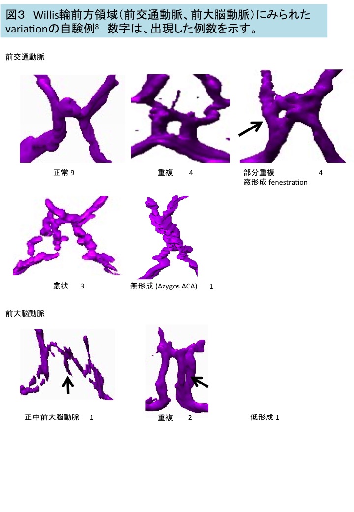
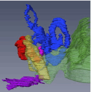
胚子期末のWillis 輪( CW )の形成を検討
先天研開設40周年記念号がAnat Recから発刊
高桑も寄稿しました
骨迷路のperiotic spaceの形成についてAnat Recに掲載
内耳形成を検討しました
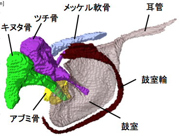
耳小骨が骨化する過程がAnat Recに掲載
耳小骨が骨化する過程を検討しました
大坂さんの修論が Anat Recに掲載
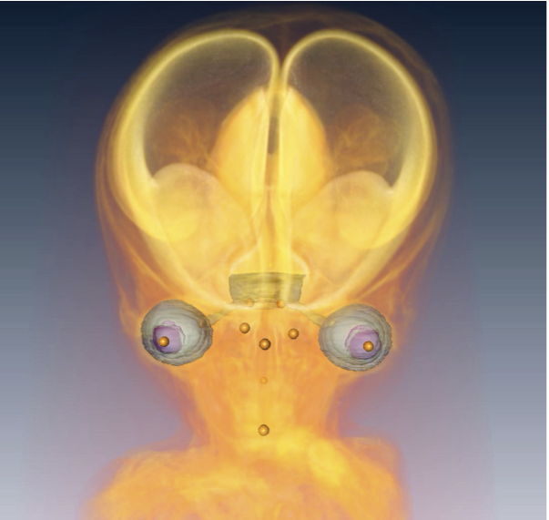
頭部・顔面形成にともなう眼の位置変化を検討しました
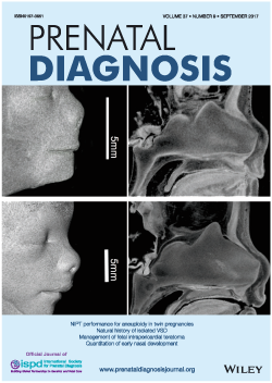
勝部先生の論文の図がprenatal Diagの表紙に採用
鼻中隔の形成について検討しました
奥村さんの卒研がPLoS Oneに掲載
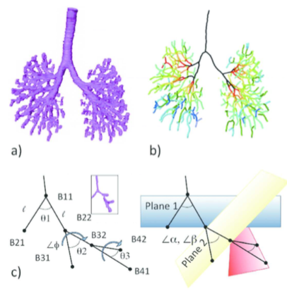
骨盤の軟骨形成期に着目し解析を進めました
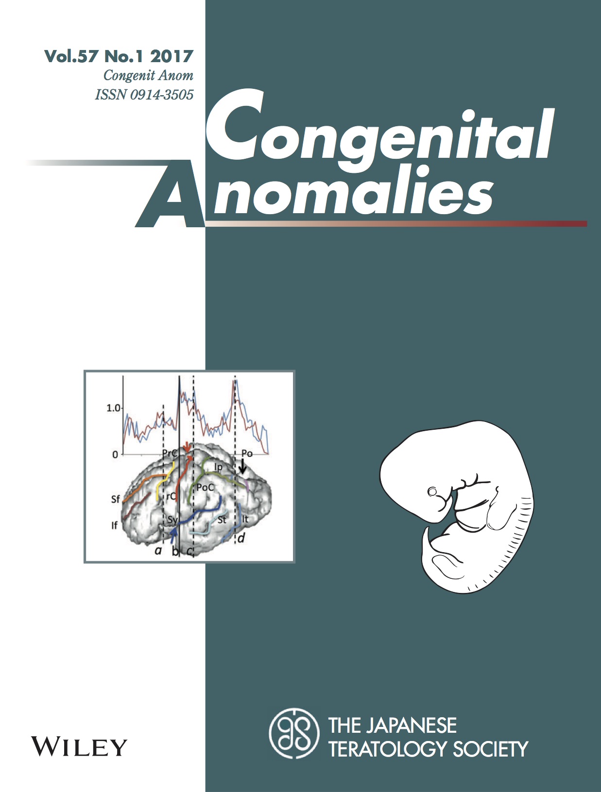
吉田さんの卒論がCongenit Anomに掲載
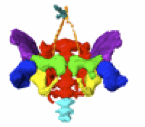
胎児の脳溝形成の様子を検討しました
Anat Recに総説を掲載
3Dデジタル胚子・胎児解析
尾関さんの修論がAnat Recに掲載
MEO の形成と外耳と内耳の接続のタイムラインを決定
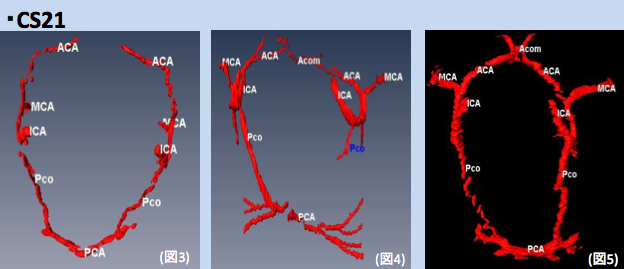
Willis輪の形成についての論文Congenit Anomに掲載
ヒト胚子期のWillis輪の形成について検討しました
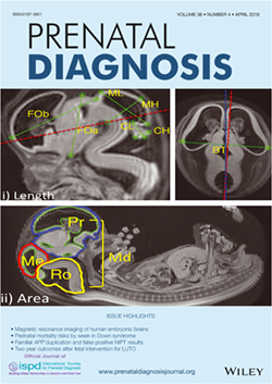
小林さんの卒論がPrenatal Diagnosisに掲載&表紙に採用
脳の発生に伴う計測値の変化、特徴を論じました