Ishikawa A, Nagai-Tanima M, Ishida K, Imai H, Yoneyama A, Yamada S, HOtani H, Aoyama T, Takakuwa T. Morphogenetic development of trochlear groove and thigh muscles from embryo to fetus in humans, PLoS One, 2025. in press.
Mechanical forces caused by fetal movements are essential for normal musculoskeletal development. However, the relationship between skeletal development and knee joint motion in utero remains unclear. We aimed to clarify the development of lower limb musculoskeletal structures during the embryonic and early fetal stages from a kinematic perspective and to compare muscle and skeletal developmental processes. We analyzed 29 human embryonic and fetal specimens. Using phase-contrast X-ray computed tomography and magnetic resonance imaging, we analyzed the morphogenesis of the knee joint components, trochlear groove angle, and hypothetical joint motion of the thigh muscles in three dimensions. The trochlear groove angle on the plane vertical to the femoral axis decreased from approximately 160° to 130° until 120 mm Crown-rump length (CRL) and remained constant until 185 mm CRL. All hypothetical joint motions increased slowly between 30 and 100 mm CRL, whereas rapidly after approximately 100 mm CRL. The protrusions of the lateral femoral condyle became more pronounced from fetal stage as they grew. The timing of the onset of fetal movement and the increase in muscle mass and joint motion during early pregnancy were consistent with those of previous studies, and the timing of the angle stabilization occurred almost simultaneously with the rapid increase in femoral muscle mass and hypothesized joint motion. It suggested that stabilization of joint morphology enables smooth joint motion, which leads to an increase in muscle mass and joint motion.
Kanahashi T, Matsubayashi J, Imai H, Otani H, Takakuwa T. Morphological Changes in the Lower Esophageal Sphincter During Early Human Fetal Development. Congenit Anom, 2025, e70032, https://doi.org/10.1111/cga.70032.
74. Hatta M, Mitsui R, Kanahashi T, Fujii S, Imai H, Otani H, Yamada S, Takakuwa T. Morphology and Morphometry of the Human Lens in the Embryonic and Early Fetal Period, Congenit Anom, 2025, 65, e70031, https://doi-org.kyoto-u.idm.oclc.org/10.1111/cga.70031.
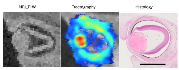
The three-dimensional structural distribution of lens fiber cells, which form an elaborate lens during the late embryonic and early fetal periods (first trimester), remains unresolved. Therefore, this study aimed to assess the sequential three-dimensional morphology, morphometry, and inner structure of human embryonic (N=10) and fetal (N=27) lenses in the first trimester using magnetic resonance imaging and tractography. The T1-weighted magnetic resonance imaging intensity and fractional anisotropy values were high at the embryonic lens, where at least two layers, the inner and outer layers, became distinguishable during the late embryonic period (Carnegie stage 21-23) and subsequent early fetal period. A comparison with histological findings indicated that the unique spherically oriented eigenvectors in the lens correspond to the lens fiber cell orientations. Furthermore, tractography demonstrated that the eigenvectors were oriented concentrically from the anterior to the posterior pole. Conversely, the eigenvectors around the centroid of the coronal section were irregular and exhibited low fractional anisotropy. Three-dimensional reconstructions, morphometry, and tractography revealed no apparent quadrant-specific deviations, as determined by the z-axis (anterior-posterior axis, AP axis), either morphologically or structurally, throughout the observation period. During the embryonic and early fetal periods, human lens cells may exhibit an exquisite hyperbolic arrangement. The ratio of transverse-to-AP length was <1 in the late embryonic period when the lens cavity was obliterated. This ratio decreased linearly from approximately 0.9 to 0.7 as the crown-rump length increased. The three-dimensional morphometry of the lens remained unaffected, irrespective of its histological structure.
73. Kumagai M, Kanahashi M, Matsubayashi J, Imai H, Otani H, Takakuwa T. Primary sulci formation in human cerebral cortex development. Anat Rec (Hoboken) 2025, 308, 3142-3156. doi: 10.1002/ar.25637
Abstract
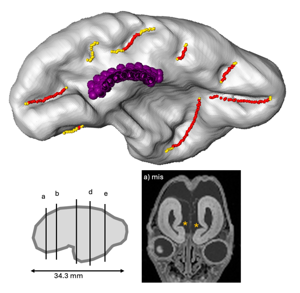
We aimed to determine the timing of appearance and the morphologic and morphometric features of the initial human cerebral sulcal formation. Using high-resolution magnetic resonance images obtained from 33 samples between 11 and 16 weeks (w) of gestation (crown-rump length <130 mm), the cerebral surface and internal structures on serial two-dimensional planes and all possible sulci on three-dimensional reconstructions were marked, allowing comparison of the positions of the sulci in the samples and inter-samples. Our method provided accurate conclusions regarding the timing of sulcal formation. Detection timing was as early as and earlier than those in previous studies using anatomical dissection and magnetic resonance imaging (MRI), respectively: <12 w for the callosum, <13 w for the hippocampal, calcarine, and parieto-occipital sulci, and <15 w for the lateral sulcus. Occasionally, an olfactory sulcus was detected. However, the cingulate sulcus could not be definitely identified. The lateral sulcus gradually appeared and changed shape. The lengths of the left and right sides of the olfactory sulci and the left side of the hippocampal sulcus increased linearly with the CRL. The length of the right side of the hippocampal sulcus and the left and right sides of the calcarine, parieto-occipital, and not determined_a sulci did not increase with the CRL The depth of the all sulci, except for the parieto-occipital sulci, increased linearly with the CRL. The sulci might not arise as if they elongate gradually but arise simultaneously over some distance. We determined the timing of the initial sulcal formation using high-resolution MRI. Our findings may significantly impact prenatal diagnosis and research on neurodevelopmental disorders.
72. Ishida, N, Kanahashi T, Matsubayashi J, Imai, H, Männer J, Yamada S, Takakuwa,T. Change in diameters of the small intestine according to embryonic and early fetal growth. J Anatomy 2025, 247, 1091-1102: 10.1111/joa.14285.
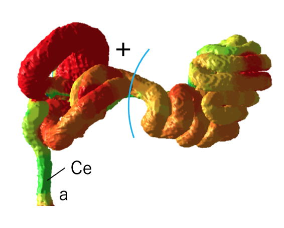
Abstract No previous studies have examined the diameter of the small intestine successively from the oral to the anal side of the small intestine. Therefore, the objectives of this study were to determine the successive intestinal diameters from the oral to the anal side (proximal to the distal) of the intestine, evaluate changes in diameter associated with growth, examine the effects of positional variation along the intestinal tract, investigate dynamic positional change from the extraembryonic coelom to the abdominal cavity, and assess the impact of complex tertiary intestinal loop formation. To this end, 14 human embryonic and fetal specimens with crown-rump lengths (CRLs) ranging from 25.6 to 69.0 mm were selected for high-resolution magnetic resonance imaging acquisition. The small intestines of the specimens were located in the extraembryonic coelom (herniation phase), transitioning phase, or abdominal cavity (return phase). The small intestine and mesentery were reconstructed in three dimensions, and the resulting morphological changes were observed and analyzed. Successive intestinal diameters from the oral to anal side of the small intestine were determined. Specifically, we observed the following: 1) gradual changes in the diameter of the position from the oral to the anal side in the jejunum-ileum, 2) the difference between the duodenum and jejunum-ileum, and 3) the difference between the superior part of the duodenum derived from the foregut and the remaining parts derived from the midgut. 4) Notably, the dynamic positional change from the extraembryonic coelom to the abdominal cavity, along with the rapid elongation and complex intestinal loop formation—a conspicuous phenomenon in the embryonic and early fetal periods—had little effect on the changes in diameter. This study indicates that increased diameter may serve as a useful indicator of intestinal development and differentiation, independent of tertiary intestinal loop formation and positional changes into and out of the abdominal cavity.
In mouse embryos, the body axis typically follows a right-handed helical pattern; however, a definitive orientation in human embryos has not been established. This study aimed to characterize the body axis orientation in human embryos (CS13–CS17) from the Kyoto Collection using MRI-based morphological assessment. Embryos were classified as right-helical (RH), left-helical (LH), and middle (M) based on morphological assessment. RH orientation was predominant at CS13, whereas it became comparable to LH at CS14. From CS15 to CS17, LH became dominant, nearly doubling the frequency of RH by CS15. The proportion of M-pattern embryos increased with advancing Carnegie Stages, reaching 70% at CS17. As vertebral column chondrification begins at CS17–18, these findings suggest that the helical body axis is established before chondrogenesis, particularly during CS13–CS15. Internal organ laterality (stomach, heart, intestines, and liver) appeared consistent among body axis orientations in CS15–CS17 embryos. The results demonstrate substantial variability in human embryonic body axis orientation, in contrast to the well-defined pattern in mice, and provide insights into body axis formation in human embryos and their potential role in left-right asymmetry.