

カテゴリー: 研究

第58回日本先天異常学会で発表: 金橋くん奨励賞受賞!
第58回日本先天異常学会で発表しました(7/27−7/29, 東京) 台風も近づき慌ただしい学会でした。 ・金橋 徹、奥村 美咲、今井 宏彦、山田 重人、高桑 徹也; 胎児期初期における骨盤形成の解析 ・鈴木 裕子、山田 […]
多元計算解剖学、第4回サマーワークショップに参加、発表
「多元計算解剖学」第4回サマーワークショップ(7月14、15日、神戸市)に参加しました。新学術領域研究「医用画像に基づく計算解剖学の多元化と高度知能化診断・治療への展開」研究班のmeetingです。今年度最終年になりまし […]
九州シンクロトロン光研究センターに行きました
佐賀県立九州シンクロトロン光研究センターに行きました。(2018.0612 位相CT撮像データからイメージ作成を行う手法を共同研究者の米山先生から習いました。
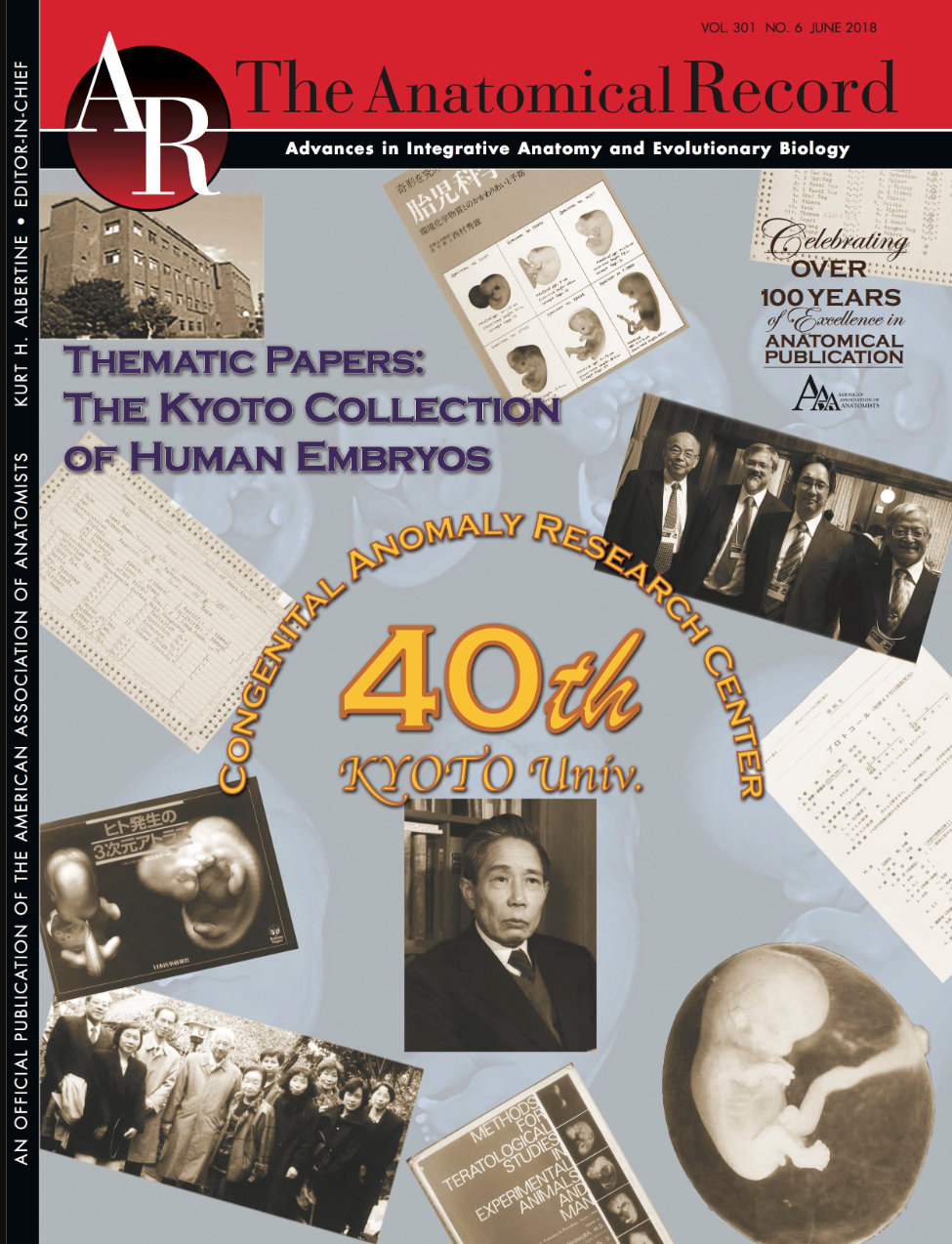
先天研開設40周年記念号がAnat Recから発刊
高桑も寄稿しました

金橋君が日本先天異常学会奨励賞を受賞しました
金橋君が2018年度日本先天異常学会奨励賞を受賞しました。おめでとうございます。 Kanahashi T, Yamada S, Tanaka M, Hirose A, Uwabe C, Kose K, Yoneyama […]
骨迷路のperiotic spaceの形成についてAnat Recに掲載
内耳形成を検討しました
多元計算解剖学第4回国際シンポジウムで発表
多元計算解剖学第4回国際シンポジウム(The 4th International Symposium on Multidisciplinary Computational Anatomy)で発表しました。(3/2-2/3 […]

2017年度; 修士論文概要
2017年度; 修士論文発表会が行われました(2018.0207; 杉浦ホール) ヒト胚子期における腎盂形成の三次元的解析 石山 華 【背景】腎盂の発生はCarnegie Stage(CS)14頃に始まる。この発生過程に […]
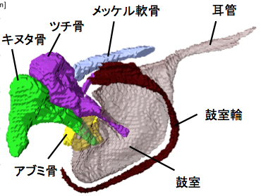
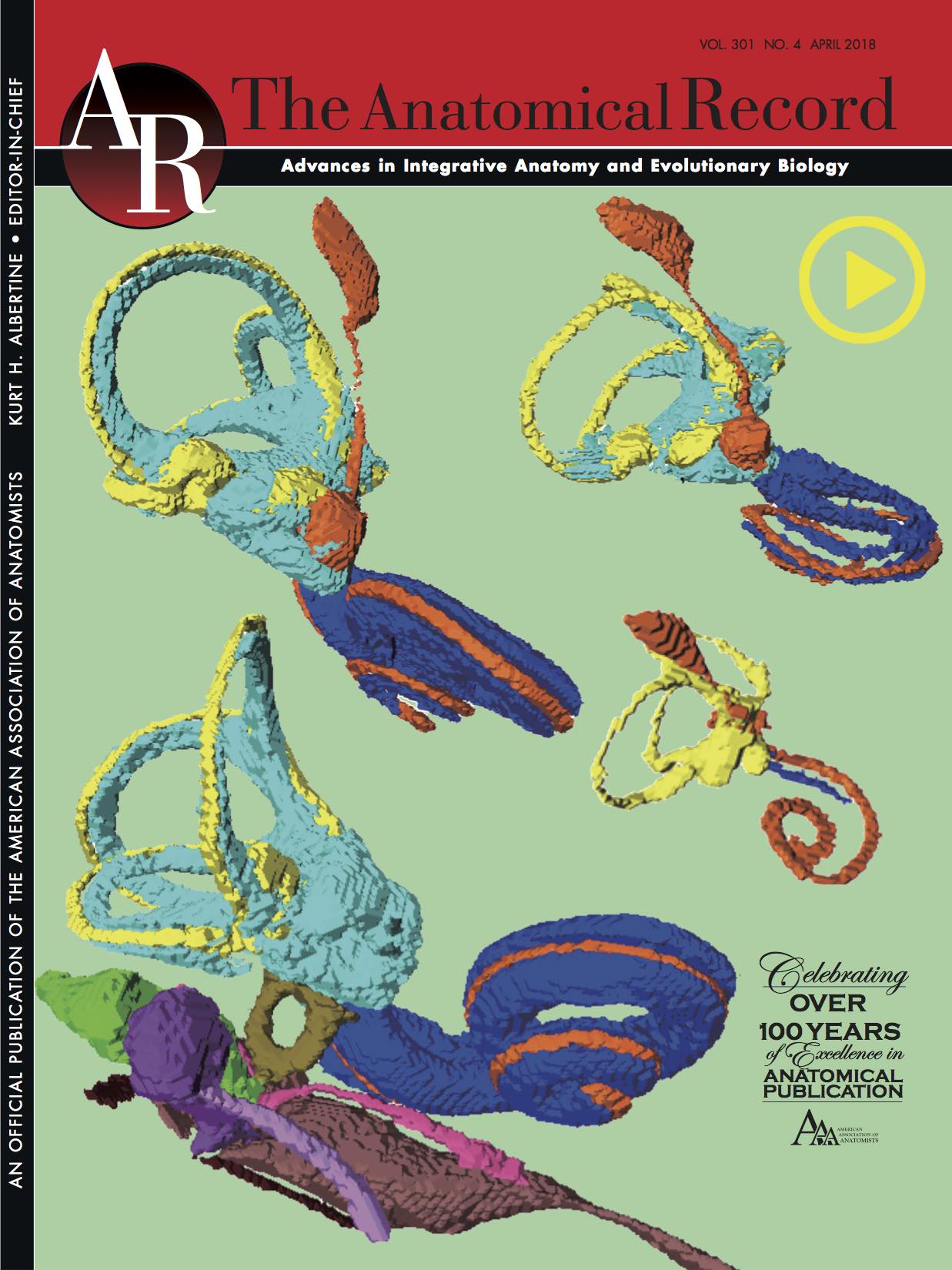
耳小骨が骨化する過程がAnat Recに掲載
耳小骨が骨化する過程を検討しました
卒業研究発表会が行われました。
2017年度;卒業研究発表会が行われました(杉浦ホール)。 ヒト胚子期における後頸部の膨隆の組織学的検討 大賀 彩子 ヒト胚子期における肋骨と胸郭の三次元的解析 奥野 香澄 (石津研) 後腎における腎小体形 […]
白石くんの博士審査会が行われました。
白石直樹くんの博士審査会が行われました。 (11月27日18時 高井ホール於) Morphology and morphometry of the human embryonic brain: A three-dimen […]
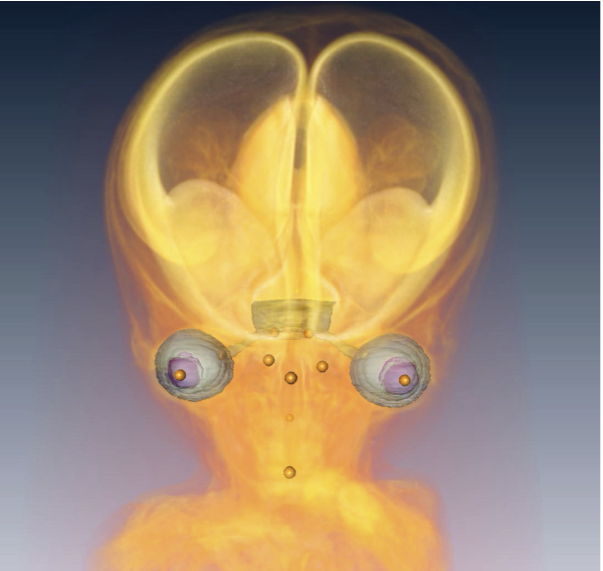
大坂さんの修論が Anat Recに掲載
頭部・顔面形成にともなう眼の位置変化を検討しました