- 最新画像技術を用いて、ヒトがどのように形作られるかの ’見える化’に取り組んでいます…
- 研究する仲間(学生・大学院生・研究者)募集中です!
|
||||
  |
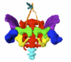
 「生理的臍帯ヘルニアはどのように腹腔に戻るのか」についての短報がCongenit Anomに受諾されました ヒトにおける生理的臍ヘルニアは、カーネギー期(CS)16~17週(5週)頃に始まる。この時期、一次中腸ループが胚外体腔へヘルニア化する。中腸は伸長し、階層的に腸ループを形成する。 ヘルニア化した中腸は、短時間で突然に腹腔へ戻ると信じられている。我々の研究を除き、復帰方法(移行期)を記述した文献は10例未満しか報告されておらず、復帰様式とメカニズムは依然として不明である。  セグメント4が臍輪に位置する標本が頻繁に観察された。これはセグメント4が臍輪を通過する際、移行速度が低下する可能性を示唆している。移行過程は、腹腔内で中腸と腸間膜のための十分な空間が確保されるまで遅くなるかもしれない。大径の盲腸は臍輪通過に時間を要する可能性があり、卵黄動脈と卵黄管の痕跡は最後に臍輪を通過する。ヘルニア期および移行期の臍帯輪断面では、近位肢と遠位肢に加え、上腸間膜動脈(SMA)および腸管枝を含む腸間膜が観察され、これらは臍帯輪内に整然と配置されていた。 80. Takakuwa T., Ishida N. How does the human herniated midgut loop return to the abdominal cavity? Congenit Anom 2025, in press.  石川さんの研究がPLoS Oneに受諾されました。 運動学的な観点から胚子期-胎児初期における下肢筋骨格構造の発達を明らかにし、筋と骨格の発達過程を比較することを目的にしました。29例の標本を位相CT, MRIを用いて得た画像から、膝関節構成要素の形態形成、滑車溝角度、および大腿筋の仮想的な関節運動を3次元で分析しました。関節形態の安定化がスムーズな関節運動を可能にし、それが筋量および関節運動の増加につながることを示唆する結果が得られました。 Ishikawa A, Nagai-Tanima M, Ishida K, Imai H, Yoneyama A, Yamada S, HOtani H, Aoyama T, Takakuwa T. Morphogenetic development of trochlear groove and thigh muscles from embryo to fetus in humans, PLoS One, 2025. in press. Mechanical forces caused by fetal movements are essential for normal musculoskeletal development. However, the relationship between skeletal development and knee joint motion in utero remains unclear. We aimed to clarify the development of lower limb musculoskeletal structures during the embryonic and early fetal stages from a kinematic perspective and to compare muscle and skeletal developmental processes. We analyzed 29 human embryonic and fetal specimens. Using phase-contrast X-ray computed tomography and magnetic resonance imaging, we analyzed the morphogenesis of the knee joint components, trochlear groove angle, and hypothetical joint motion of the thigh muscles in three dimensions. The trochlear groove angle on the plane vertical to the femoral axis decreased from approximately 160° to 130° until 120 mm Crown-rump length (CRL) and remained constant until 185 mm CRL. All hypothetical joint motions increased slowly between 30 and 100 mm CRL, whereas rapidly after approximately 100 mm CRL. The protrusions of the lateral femoral condyle became more pronounced from fetal stage as they grew. The timing of the onset of fetal movement and the increase in muscle mass and joint motion during early pregnancy were consistent with those of previous studies, and the timing of the angle stabilization occurred almost simultaneously with the rapid increase in femoral muscle mass and hypothesized joint motion. It suggested that stabilization of joint morphology enables smooth joint motion, which leads to an increase in muscle mass and joint motion.  第24回日本心臓血管発生研究会で発表しました(2025.11.28-29, 淡路島) 藤井瀬菜, 磯谷菜穂子,今井宏彦, 米山明男, 山田重人, 高桑徹也:ヒト胚子期および胎児期における静脈管の三次元的解析と血流シミュレー ションの検討 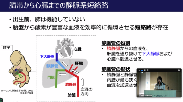  従来、下部食道括約筋 (lower esophageal sphincter, LES)は、成人では横隔膜の脚部と重なって位置する「生理的括約部」として知られていますが、その胎児期における発達過程は十分に明らかにされていません。本研究では、ヒト胎児期におけるLESの形成過程を、7テスラMRIを用いた三次元解析により詳細に検討しました。受精後9〜13週相当(頭殿長34〜103 mm)の24体を対象に、食道下端(LES部)とその頭側領域における外径および内腔径を比較した結果、特にLES部では内腔が相対的に狭くなり、壁の肥厚が顕著であることが明らかになりました。これらの変化はおおよそ頭殿長70 mm(10週頃)から認められ、形態的成熟が機能的成熟に先行することが示唆されます。 Kanahashi T, Matsubayashi J, Imai H, Otani H, Takakuwa T. Morphological Changes in the Lower Esophageal Sphincter During Early Human Fetal Development. Congenit Anom, 2025, e70032, https://doi.org/10.1111/cga.70032. ヒト胚子、胎児の水晶体形成についての論文がCongenital Anomaliesに受諾、掲載(電子版)されました。三ツ井さんの卒業研究を八田さんが引き継いで発展させました。 水晶体は胚子期末は球体に近いのですが、徐々に双曲線状の楕円に変わっていきます。水晶体線維細胞という細胞の集合体で形成されますが、みごとに歪みのない形を維持しながら成長します。
74. Hatta M, Mitsui R, Kanahashi T, Fujii S, Imai H, Otani H, Yamada S, Takakuwa T. Morphology and Morphometry of the Human Lens in the Embryonic and Early Fetal Period, Congenit Anom, 2025, 65, e70031, https://doi-org.kyoto-u.idm.oclc.org/10.1111/cga.70031. 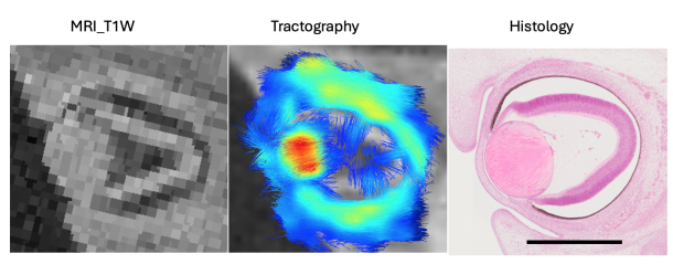 The three-dimensional structural distribution of lens fiber cells, which form an elaborate lens during the late embryonic and early fetal periods (first trimester), remains unresolved. Therefore, this study aimed to assess the sequential three-dimensional morphology, morphometry, and inner structure of human embryonic (N=10) and fetal (N=27) lenses in the first trimester using magnetic resonance imaging and tractography. The T1-weighted magnetic resonance imaging intensity and fractional anisotropy values were high at the embryonic lens, where at least two layers, the inner and outer layers, became distinguishable during the late embryonic period (Carnegie stage 21-23) and subsequent early fetal period. A comparison with histological findings indicated that the unique spherically oriented eigenvectors in the lens correspond to the lens fiber cell orientations. Furthermore, tractography demonstrated that the eigenvectors were oriented concentrically from the anterior to the posterior pole. Conversely, the eigenvectors around the centroid of the coronal section were irregular and exhibited low fractional anisotropy. Three-dimensional reconstructions, morphometry, and tractography revealed no apparent quadrant-specific deviations, as determined by the z-axis (anterior-posterior axis, AP axis), either morphologically or structurally, throughout the observation period. During the embryonic and early fetal periods, human lens cells may exhibit an exquisite hyperbolic arrangement. The ratio of transverse-to-AP length was <1 in the late embryonic period when the lens cavity was obliterated. This ratio decreased linearly from approximately 0.9 to 0.7 as the crown-rump length increased. The three-dimensional morphometry of the lens remained unaffected, irrespective of its histological structure. 熊谷さんの卒業研究がAnat Recに受諾されました。1次脳溝(脳のしわ)が、どのように最初できてくるかを検討しました。 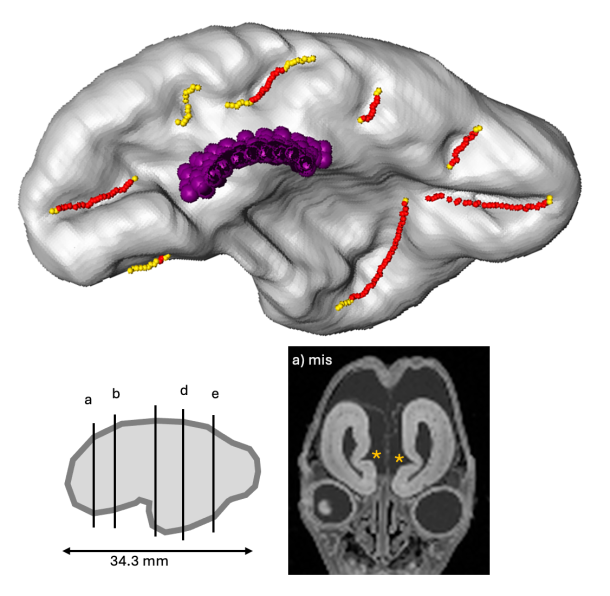
73. Kumagai M, Kanahashi M, Matsubayashi J, Imai H, Otani H, Takakuwa T. Primary sulci formation in human cerebral cortex development. Anat Rec (Hoboken) 2025, 308, 3142-3156. doi: 10.1002/ar.25637 Abstract We aimed to determine the timing of appearance and the morphologic and morphometric features of the initial human cerebral sulcal formation. Using high-resolution magnetic resonance images obtained from 33 samples between 11 and 16 weeks (w) of gestation (crown-rump length <130 mm), the cerebral surface and internal structures on serial two-dimensional planes and all possible sulci on three-dimensional reconstructions were marked, allowing comparison of the positions of the sulci in the samples and inter-samples. Our method provided accurate conclusions regarding the timing of sulcal formation. Detection timing was as early as and earlier than those in previous studies using anatomical dissection and magnetic resonance imaging (MRI), respectively: <12 w for the callosum, <13 w for the hippocampal, calcarine, and parieto-occipital sulci, and <15 w for the lateral sulcus. Occasionally, an olfactory sulcus was detected. However, the cingulate sulcus could not be definitely identified. The lateral sulcus gradually appeared and changed shape. The lengths of the left and right sides of the olfactory sulci and the left side of the hippocampal sulcus increased linearly with the CRL. The length of the right side of the hippocampal sulcus and the left and right sides of the calcarine, parieto-occipital, and not determined_a sulci did not increase with the CRL The depth of the all sulci, except for the parieto-occipital sulci, increased linearly with the CRL. The sulci might not arise as if they elongate gradually but arise simultaneously over some distance. We determined the timing of the initial sulcal formation using high-resolution MRI. Our findings may significantly impact prenatal diagnosis and research on neurodevelopmental disorders. このたび、以前から取り組んできた「教育用寄生虫標本のデジタル化および共有活動」が高く評価され、表彰されることとなりました。 本活動は、寄生虫学教育の質向上と教材のアクセシビリティ向上を目的としており、今後も教育現場や研究機関との連携を通じて、標本の有効活用と知識の普及に努めてまいります。   第79回日本人類学会で発表しました (2025.10.11-13: 下関) 金橋 徹、松林 潤、今井 宏彦、山田 重人、大谷 浩 、高桑 徹也: ヒト胎児骨盤傾斜角の性差の検討 青江 春菜、松林 潤、藤井 瀬菜、金橋 徹、今井 宏彦、大谷 浩、山田 重人、高桑 徹也: ヒト胎児期初期における歯胚の三次元解析 熊谷 美優、金橋 徹、藤井瀬菜、今井 宏彦、大谷 浩、多賀 厳太郎、山田重人、高桑 徹也: ヒト胎児における大脳基底核原基の形成過程の検討 倭 友希、田村 祥太郎、松田 幸樹、松林 潤、藤井 瀬菜、金橋 徹、今井 宏彦、 米山 明男、山田 重人、高桑 徹也: 胚子期から胎児期初期にかけてのヒトの足部の形態形成  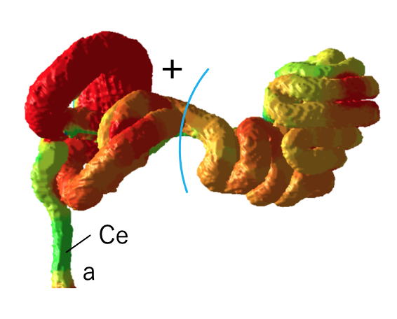 石田さんの修士論文がJ Anatomyに受諾されました。胚子期から胎児期初期の中腸の形成に伴う太さについて検討しました。口側から肛門側へと太さが漸減する様子は美しいです。 72. Ishida, N, Kanahashi T, Matsubayashi J, Imai, H, Männer J, Yamada S, Takakuwa,T. Change in diameters of the small intestine according to embryonic and early fetal growth. J Anatomy 2025, 247, 1091-1102: 10.1111/joa.14285.
Abstract |
|
||