


八田さんの修士論文がCongenit Anomに掲載
ヒト胚子、胎児の水晶体形成についての論文がCongenital Anomaliesに受諾、掲載(電子版)されました。三ツ井さんの卒業研究を八田さんが引き継いで発展させました。
水晶体は胚子期末は球体に近いのですが、徐々に双曲線状の楕円に変わっていきます。水晶体線維細胞という細胞の集合体で形成されますが、みごとに歪みのない形を維持しながら成長します。
- MRI, DTIを用いて、ヒト胚子期後期から胎児期初期の水晶体の三次元形態、形態計測、および内部構造を経時的に評価
- T1強調MRI, DTIでは、胚子期末から水晶体は内外層の2層が識別可能
- 水晶体特有の球面状の固有ベクトルは、水晶体線維細胞の配向と一致
- Tractographyでは、固有ベクトルが前極から後極にかけて同心円状に配向
- 冠状断面重心周囲の固有ベクトルは不規則で、異方性率が低い
- 胚子期後期から胎児期初期には、ヒトの水晶体細胞は精巧な双曲配置を示す
- 胎生期後期には、横方向/AP方向の比は1未満
- 頭殿長が増加するにつれて、約0.9から0.7に直線的に減少
74. Hatta M, Mitsui R, Kanahashi T, Fujii S, Imai H, Otani H, Yamada S, Takakuwa T. Morphology and Morphometry of the Human Lens in the Embryonic and Early Fetal Period, Congenit Anom, 2025, 65, e70031, https://doi-org.kyoto-u.idm.oclc.org/10.1111/cga.70031.
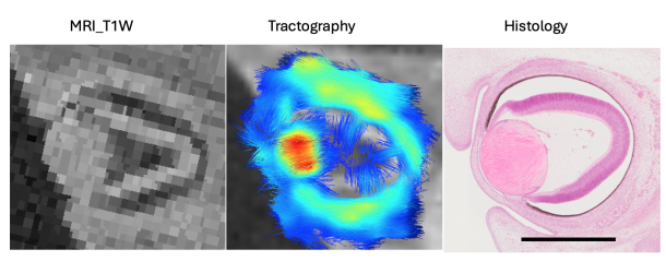
The three-dimensional structural distribution of lens fiber cells, which form an elaborate lens during the late embryonic and early fetal periods (first trimester), remains unresolved. Therefore, this study aimed to assess the sequential three-dimensional morphology, morphometry, and inner structure of human embryonic (N=10) and fetal (N=27) lenses in the first trimester using magnetic resonance imaging and tractography. The T1-weighted magnetic resonance imaging intensity and fractional anisotropy values were high at the embryonic lens, where at least two layers, the inner and outer layers, became distinguishable during the late embryonic period (Carnegie stage 21-23) and subsequent early fetal period. A comparison with histological findings indicated that the unique spherically oriented eigenvectors in the lens correspond to the lens fiber cell orientations. Furthermore, tractography demonstrated that the eigenvectors were oriented concentrically from the anterior to the posterior pole. Conversely, the eigenvectors around the centroid of the coronal section were irregular and exhibited low fractional anisotropy. Three-dimensional reconstructions, morphometry, and tractography revealed no apparent quadrant-specific deviations, as determined by the z-axis (anterior-posterior axis, AP axis), either morphologically or structurally, throughout the observation period. During the embryonic and early fetal periods, human lens cells may exhibit an exquisite hyperbolic arrangement. The ratio of transverse-to-AP length was <1 in the late embryonic period when the lens cavity was obliterated. This ratio decreased linearly from approximately 0.9 to 0.7 as the crown-rump length increased. The three-dimensional morphometry of the lens remained unaffected, irrespective of its histological structure.
Related Posts

上腸間膜動脈の形成についての論文が掲載

石川さんの研究がPLoS Oneに掲載
