


Marieさんの論文がPLoS Oneに掲載されました。PLoS Oneは査読が早い印象がありましたが、今回の初回の査読は7ヶ月もかかりました。読んでくれる研究者がみつからなかったのでしょうか。
Marieさんの博士論文として申請、審査を受ける予定です。
Saizonou MA, Kitazawa H, Kanahashi T, Yamada S, Takakuwa T, Epithelial development of the urinary collecting system in the human embryo, PLOS ONE 19(4): e0301778. https://doi.org/10.1371/journal.pone.0301778
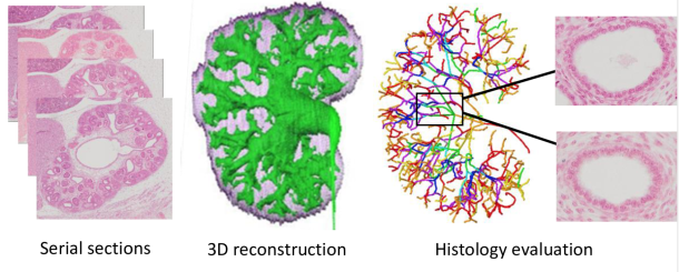
連続組織切片画像とそれにもとずいて作成した立体像のデータセットを活用して、立体像から尿路の分岐を把握し分岐次数ごとにどのような組織学的形態を示すかを、組織切片に戻って検討しました。
Abstract
The urinary collecting system (UCS) consists of organized ducts that collect urine from the nephrons and transport it to the ureter and bladder. Understanding the histogenesis of the UCS is critical. Thirty human embryos between the Carnegie stages (CS) 18 and 23 were selected from the Congenital Anomaly Research Center, Kyoto, Japan. Epithelia of the UCS, ureter, and bladder of each sample were randomly selected. Histological findings of the epithelia were analyzed according to the following criteria: type of epithelium, presence or absence of glycogen, percentage of migrated nuclei, percentage of cells in mitosis, and the surrounding mesenchyme. A thickened epithelium lining a narrow luminal cavity was observed in the pre-expanded pelvic specimens at CS18-CS23. At CS23, after pelvic expansion, the UCS showed a thin epithelium with a large luminal cavity mainly located on the early branches, whereas the epithelium covering the subsequent branches had medium thickness. Histological characteristics differed depending on the UCS part and sample stage. The degree of differentiation was evaluated, revealing that in CS18-CS23 pre-expanded pelvis specimens, the undifferentiated epithelium was found in the zeroth to third/fifth generation, whereas at CS23, after pelvic expansion, a differentiated epithelium covered the UCS zeroth to seventh generation. In a comparison of the urothelial epithelium between the UCS, ureter, and bladder, we found that urinary tract differentiation may be initiated in the bladder, followed by the ureter, UCS zeroth to seventh generations, and finally, UCS eighth to end generations. An understanding of the histogenesis of embryonic stage UCS can aid in the clinical management of congenital urinary tract defects and other diseases.
集合管(UCS)は、ネフロンから尿を集め、尿管と膀胱へ送る管路から構成されています。UCSの組織発生を理解することは非常に重要です。京都大学先天異常研究センターから、カーネギーステージ(CS)18~23のヒト胎児30例を採取しました。各標本において、UCS、尿管、膀胱の上皮を無作為に抽出しました。上皮の組織学的所見は、上皮の種類、グリコーゲンの有無、遊走核の割合、有糸分裂中の細胞の割合、および周囲の間葉系という基準に基づいて分析しました。拡張前の骨盤標本において、CS18~CS23の狭い管腔を裏打ちする肥厚した上皮が観察されました。骨盤拡張後のCS23では、UCSは主に初期の枝に位置する大きな管腔を持つ薄い上皮を示し、それ以後の枝を覆う上皮は中程度の厚さであった。組織学的特徴は、UCSの部位と標本の段階によって異なった。分化の程度を評価したところ、拡張前のCS18-CS23骨盤標本では、第0世代から第3/5世代に未分化上皮が見られたのに対し、骨盤拡張後のCS23では、第0世代から第7世代のUCSが分化上皮で覆われていることが明らかになった。UCS、尿管、膀胱の尿路上皮を比較すると、尿路分化は膀胱で開始され、続いて尿管、第0世代から第7世代のUCS、そして最終的に第8世代から末世代のUCSへと進むことがわかった。胎児期の UCS の組織発生を理解することは、先天性尿路欠損症やその他の疾患の臨床管理に役立ちます。