

カテゴリー: 活動記録
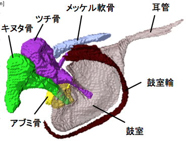
耳小骨が骨化する過程がAnat Recに掲載
耳小骨が骨化する過程を検討しました
卒業研究発表会が行われました。
2017年度;卒業研究発表会が行われました(杉浦ホール)。 ヒト胚子期における後頸部の膨隆の組織学的検討 大賀 彩子 ヒト胚子期における肋骨と胸郭の三次元的解析 奥野 香澄 (石津研) 後腎における腎小体形 […]
白石くんの博士審査会が行われました。
白石直樹くんの博士審査会が行われました。 (11月27日18時 高井ホール於) Morphology and morphometry of the human embryonic brain: A three-dimen […]
大坂さんの修論が Anat Recに掲載
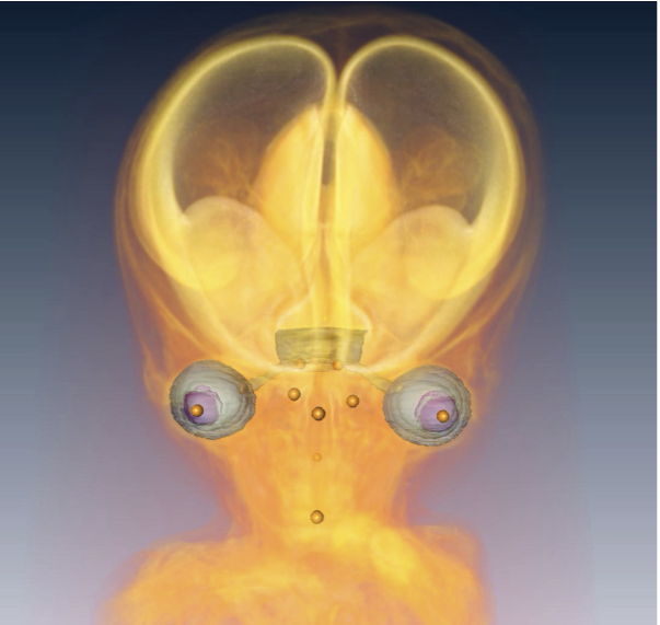
頭部・顔面形成にともなう眼の位置変化を検討しました
多元計算解剖学第3回サマーワークショップ参加
「多元計算解剖学」第3回サマーワークショップ(9月26、27日、神戸市)に参加しました。新学術領域研究「医用画像に基づく計算解剖学の多元化と高度知能化診断・治療への展開」研究班のmeetingです。
ゲッチンゲン大学に標本の撮像に行きました。
ゲッチンゲン大学にガラス標本のスキャンに行ってきました。 先天異常解析センター、ゲッチンゲン大学解剖学研究室との共同研究で、今年で3年目です。 今年度は9月の2-4週にのべ8名、本研究室からはM1の鈴木さん、西谷さんが参 […]
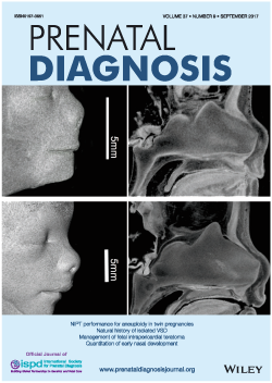
勝部先生の論文の図がprenatal Diagの表紙に採用
鼻中隔の形成について検討しました
第57回先天異常学会で発表
57回日本先天異常学会学術集会・第6回日本DOHaD学会 合同学術集会 ( 2017.8.26-28, 東京, 新宿)で発表しました。 ヒト胚子期~胎児期初期における大腿骨の形態形成の解析;鈴木 裕子、山田 重人、上部千 […]

第106回日本病理学会で発表:高石くん日本病理学会総会最優秀賞受賞!
第106回日本病理学会で発表しました。(2017.4.27-29、新宿) 学生部門で高石くんが日本病理学会総会最優秀賞を受賞しました。 英語口演 □ ヒト気管支発生の 3 次元的解析 Three-dimensional […]
多元計算解剖学キックオフシンポジウム
平成29年度 新学術領域研究「多元計算解剖学」シンポジウム(4.21(金)10時〜@名大) 高桑は、スポーツ実習、細胞組織学実習等、終日実習をする日程になっていましたので、修士2年石山さんが代わりに発表しました。 石山さ […]
多元計算解剖学・第3回 国際シンポジウム
新学術領域研究「多元計算解剖学」第3回 国際シンポジウム(2017.3.8-9,奈良県文化会館) で発表しました。 A01-KB004 Three-dimensional Analysis of the Bronchia […]
奥村さんの卒研がPLoS Oneに掲載
骨盤の軟骨形成期に着目し解析を進めました