

カテゴリー: 原著論文・総説
上野さんの修論がAnat Recに掲載
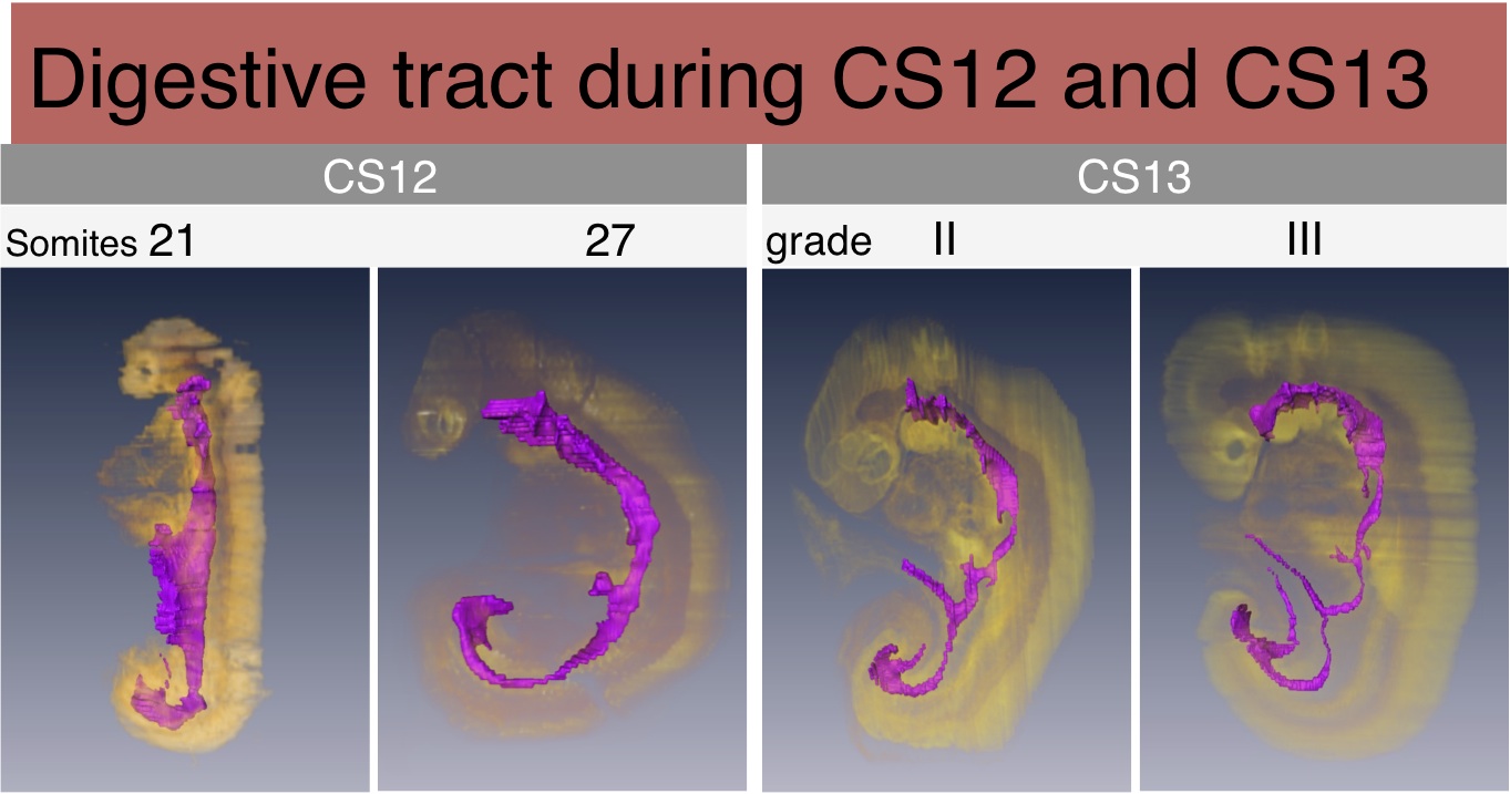
Somite stageヒト胚において消化管由来原基の形態と分化のTimeLineを検討
尾関さんの修論が Congenit Anomに掲載
500例以上の外耳観察の成果です
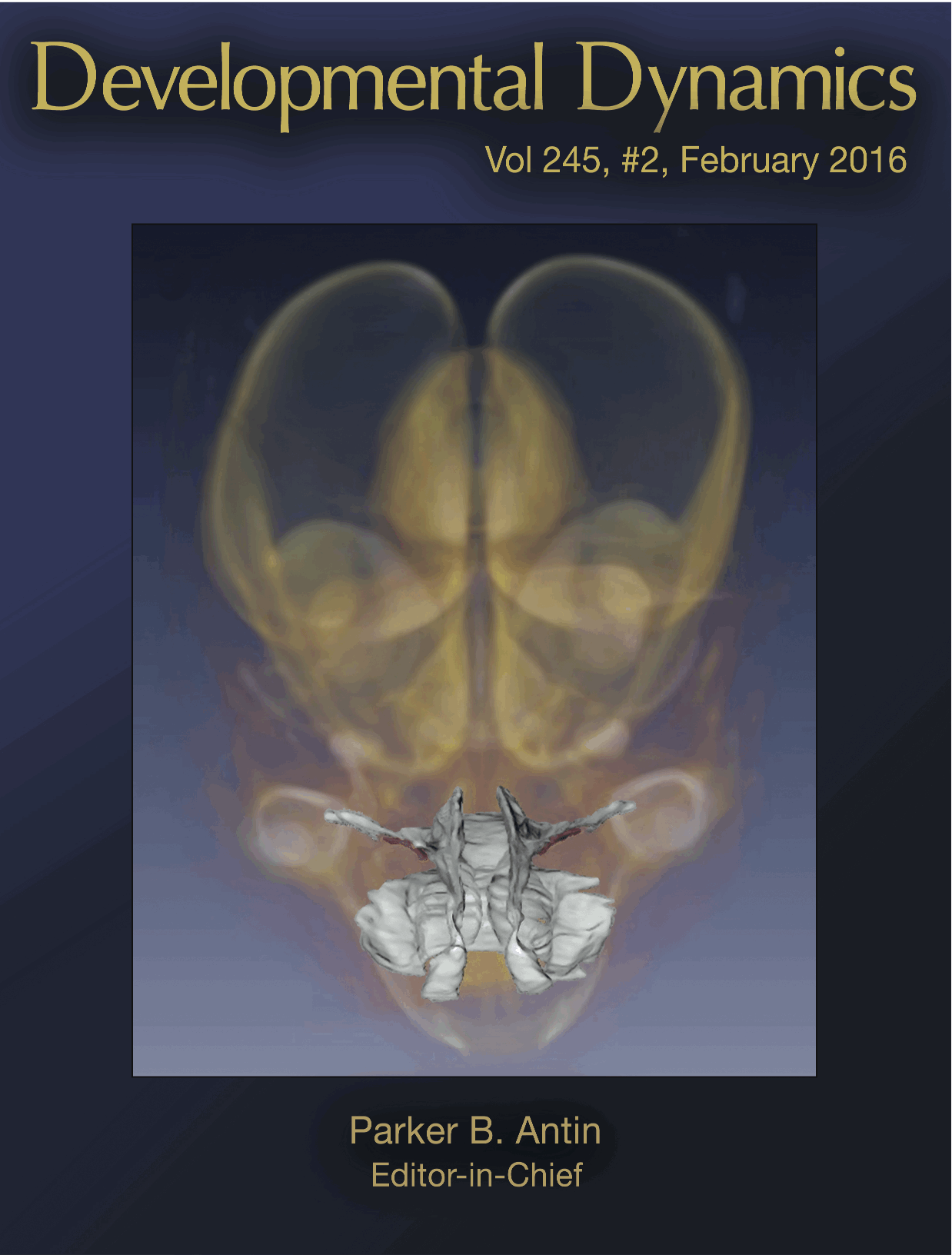
金橋君の描いた図がDevelopmental Dynamicsの表紙に採用
ヒト胎児の咽頭口蓋領域の解析
植田さんの卒業研究が Anat Recに掲載
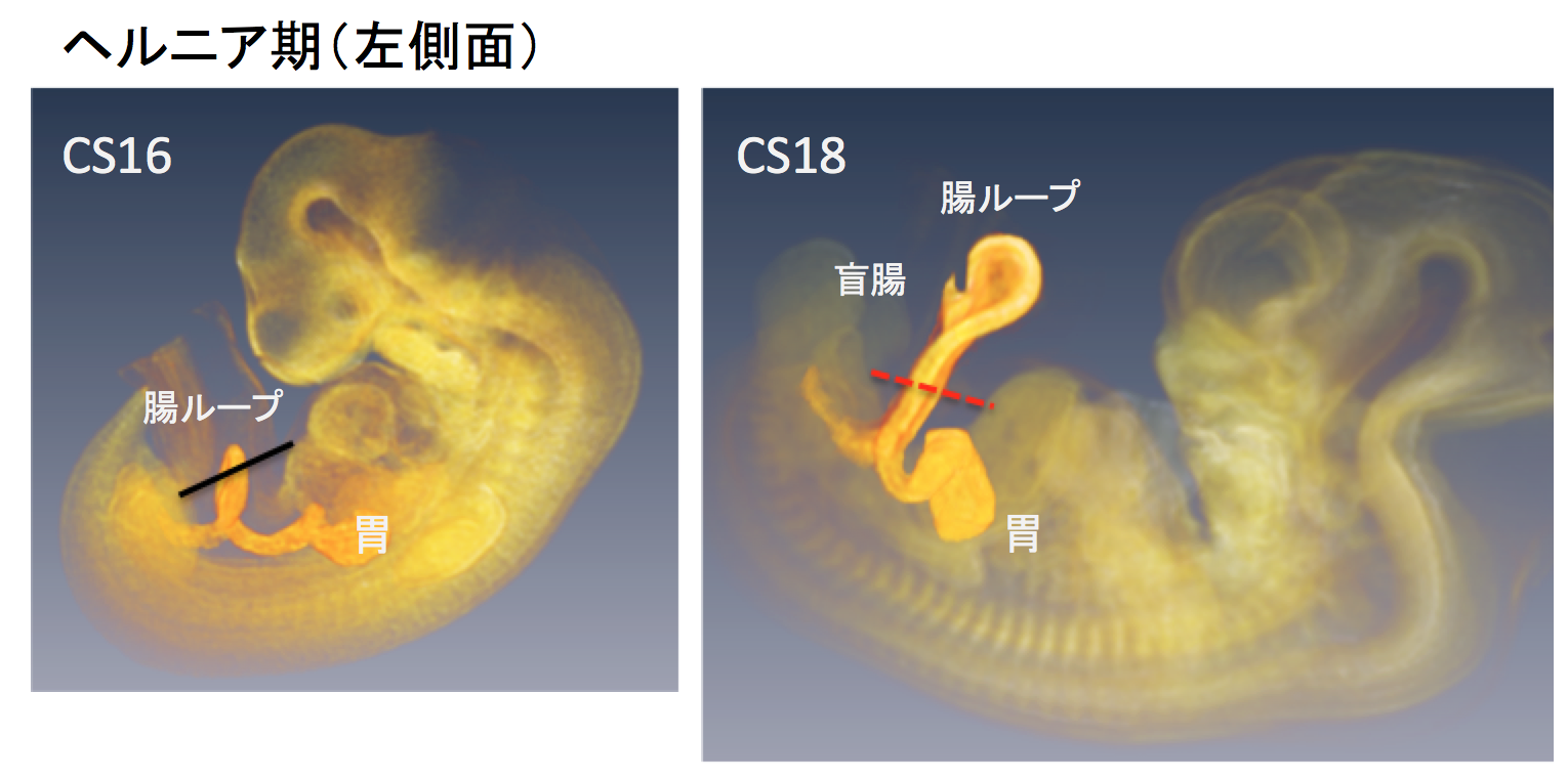
ヒト胚子期の中腸の回転と臍帯内への生理的ヘルニアについて検討しました。
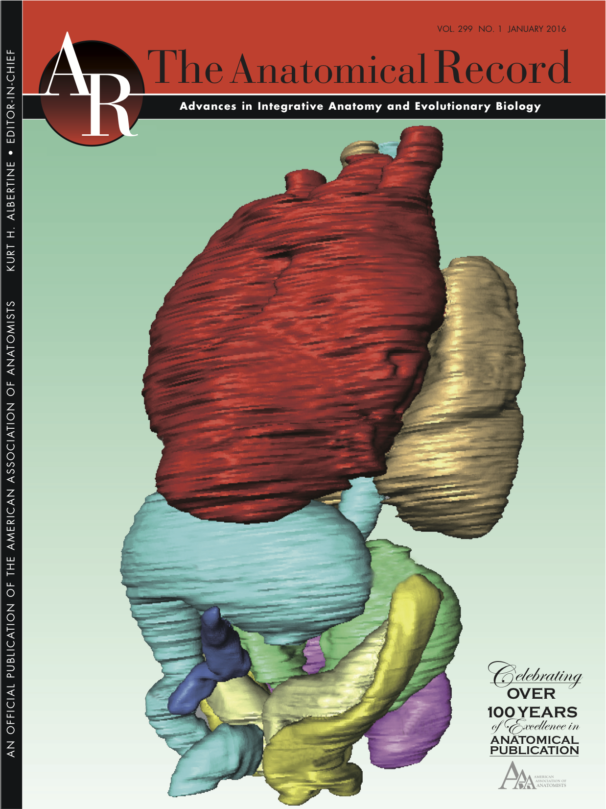
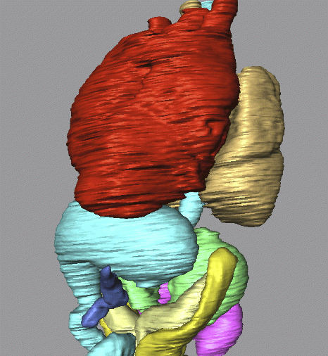
Anat Rec 299巻1号の表紙に肝形成不全例が採用
肝形成不全例の解析
豊田さんの卒業研究がAnat Recに掲載
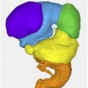
内耳(膜迷路)の形態形成を検討
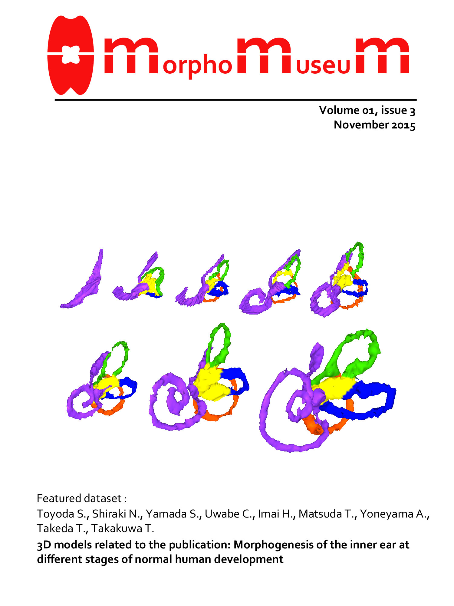
内耳・膜迷路の3D画像が MorphoMuseuMの表紙に採用
ヒト胚子の内耳・膜迷路の3D画像
総説”Human embryology”が発行
ヒトの発生学についての総説が発行されました。New Discoveries in Embryologyという書籍の第5章に収載されています。internetからdownloadできます。 Yamada S, Hill M […]
金橋君の修論がAnat Recに掲載
CS18 -21 の肝形成不全の有病率は、約 1.7%
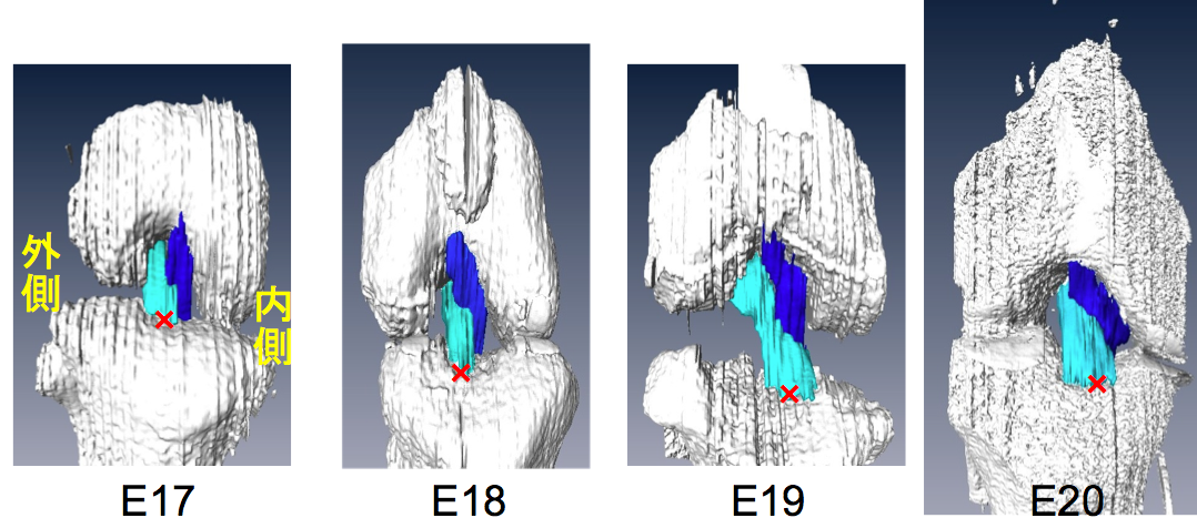
脳の形態形成の3次元データがMorphoMuseuMに掲載
胚子期の脳、脳室の形態像に関する3次元データ
張・高石・樋口君の論文が PlosOneに掲載
ラットの膝関節のACL, PCL靭帯の形成を検討
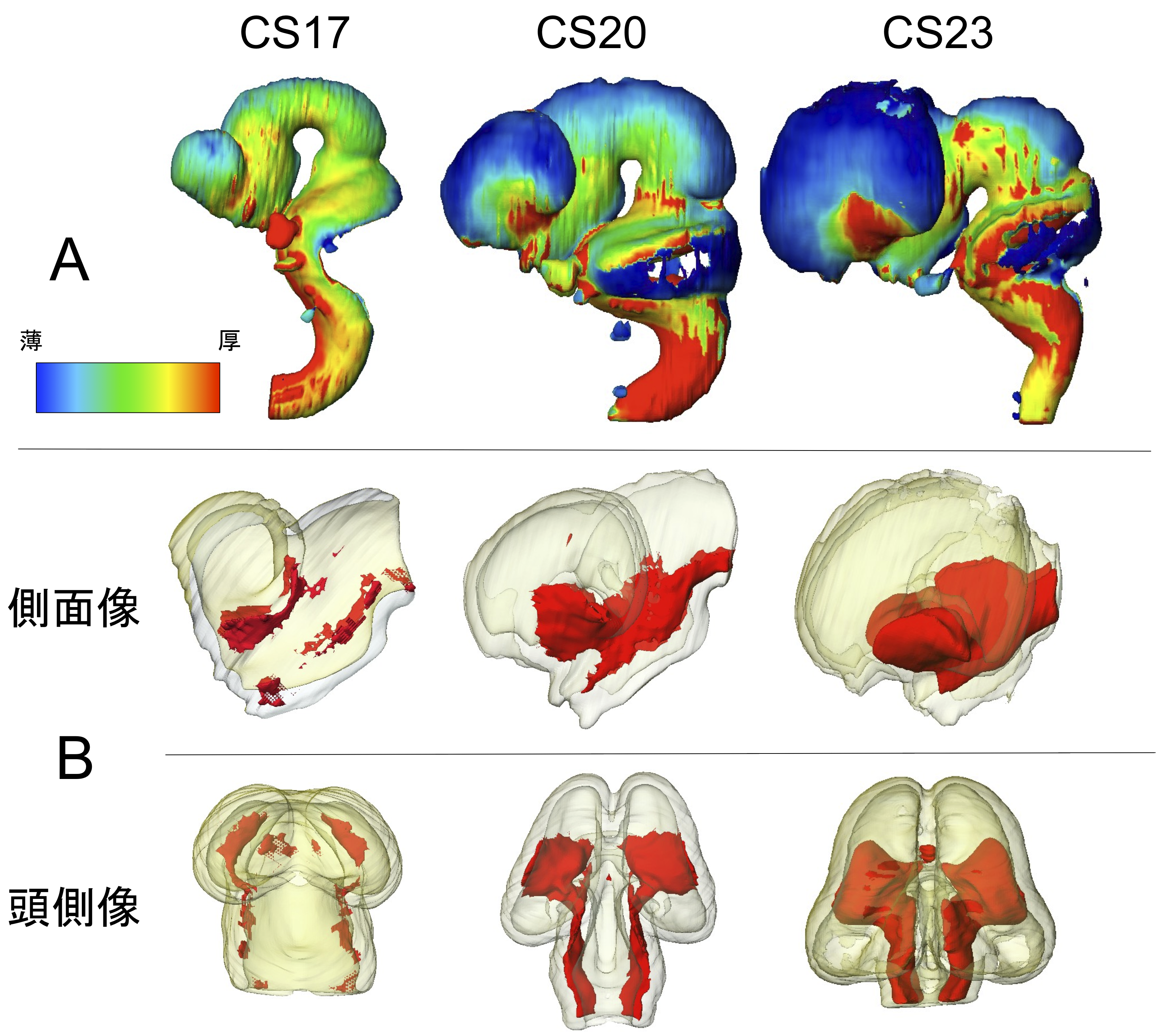
ヒト胚子期における脳形態形成の解析, NeuroImage, Data in Briefに掲載
ヒト胚子期の脳の三次元形成を提示