アクセスカウンター

2025年7月
 123456
78910111213
14151617181920
21222324252627
28293031  

第61回日本先天異常学会で発表

第61回日本先天異常学会で発表しました。(2021.08.7-8, 東京, on line開催)

拡散テンソルイメージングを応用したヒト胎児の横隔膜形成過程の解析; 金橋 徹、今井宏彦、大谷浩、高桑徹也

ヒト胚子期における気管支樹のvariationの検討;藤井 瀬菜、村中太河、松林潤、山田重人、米山明男、高桑徹也

藤井さんが学会奨励賞を受賞いたしました。おめでとうございます。

受賞講演要旨

  • 【背景・目的】多くの成人肺の解剖学的研究が、区域気管支の分岐構造に関するvariationを報告している。この形態学的差異が生じる時期は明らかでない。本研究は、ヒト胚子の気管支樹の形態変化を観察し、気管支のvariationが生じる時期を検討した。
  • 【対象・方法】京都コレクション保有のカーネギーステージ(CS)15~23のヒト胚子標本計48体を対象とした。全個体の位相 CT 画像を取得し、画像処理ソフトウェア Amira を用いて気管支樹を抽出し立体像を作成した。
  • 【結果】CS15~23 において、気管と葉気管支の形態に個体差は認めなかった。葉気管支はCS15, 16にて形成されていた。左右の一次気管支芽上の対称な位置に右中葉と左上葉を形成している個体と、さらに右上葉を形成している個体を認めた。区域気管支と亜区域気管支は、CS17~19 にかけて形成が進んでおり、同一ステージの個体間にて形成の程度にばらつきが生じていた。CS20~23の気管支樹における区域気管支の形態として、左右の上葉においてそれぞれ4種類、右中葉と左右下葉ではそれぞれ2種類の計14種を同定した。
  • 【考察】観察した全個体にて、気管と葉気管支は成人の気管支樹と同様の構造を示した。この結果は、葉気管支までは個体に関わらず典型的な構造を形成することを示唆している。CS15~19の気管支樹の観察から、葉気管支は一定の順序で形成される可能性があること、区域気管支の形成速度には個体差があることが明らかになった。さらに、CS20~23で同定した区域気管支の形態は、成人の気管支においても同様の形態が報告されていた。この結果は、成人の気管支樹の形態のvariationが胚子期において決定し、生涯にわたって継続する可能性を示唆している。本研究は医の倫理委員会で承認されている。(R0316)

第2回医薬系研究交流サロンで発表

第2回医薬系研究交流サロンで発表しました。(2021年4月19日 (月)~21日(水)17:00~19:00,  オンライン)

Jou Ting Huang;Fixation of the colorectum to the retroperitoneum during human fetal development

掛谷真樹;The quantitative analysis of returning phase of intestine in physiological umbilical herniation 口頭発表もしました。

福井成美;ヒト胎児期初期における左心房形態形成~左心耳の形成と肺静脈が左心房に取り込まれる過程~
磯谷菜穂子;ヒト胚子期・胎児期初期における静脈管の定量的形態解析

石田かのん;ラット膝関節の発生と半月大腿靭帯の形態形成

金橋 徹;ヒト胎児期初期における横隔膜形成過程の解析

コロナ感染症もあり、on lineで開催されました。


第25回日本基礎理学療法学会学術大会で発表

第25回日本基礎理学療法学会学術大会で発表しました。2020.12.12-13 @仙台市(on line開催)


石川葵、田中麻衣、杉山寛恵、池口良輔、高井治美、鳥井蓉子、國富芳博、秋枝静香、谷間桃子、青山朋樹: 神経様3次元組織体の生死判定技術の検討

石田かのん、谷間桃子、石川葵、高石亮太、高桑徹也、青山朋樹: ラット膝関節の発生と 半月大腿靭帯の形態形成

第60回日本先天異常学会で発表

第60回日本先天異常学会で発表しました。(2020.07.11-12, 神戸)

新型コロナウイルス感染症の影響でon line 学術開催となりました。

金橋 徹、山田重人、米山明男、高桑徹也, ヒト胚子期に起こる生理的臍帯ヘルニアの発生要因‐肝形成との関連に基づいた形態及び形態計測学的検討

藤井 瀬菜, 村中 太河, 松林 潤, 米山 明男, 兵藤 一行, 山田 重人, 高桑 徹也, ヒト胚子期における気管支の非対称性の定量的検討

共同研究者の勝部先生、東島先生が奨励賞を受賞しました。おめでとうございます。

勝部 元紀 先生
Critical Growth Processes for the Midfacial Morphogenesis in the Early Prenatal Period
東島 沙弥佳 先生
Tail reduction process during human embryonic development

第109回日本病理学会総会で発表

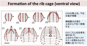
第109回日本病理学会総会で発表しました (2020.07.01-07.31, 福岡(オンライン))

高桑徹也、山田重人、米山明男. ヒト胚子肋骨の形態形成は、共通の肋骨の形状変化のどのあたりに相当するかという尺度で示すことができる

第125回日本解剖学会で発表

第125回日本解剖学会で発表しました(3/25-3/27, 山口)

高桑徹也、寺島芽衣、石川葵、山田重人: ヒト胚子期終期における大脳層構造の三次元的解析

藤井瀬菜、村中 太河、松林 潤、米山明男、兵藤一行、山田重人、高桑徹也:ヒト胚子期における気管支形成の定量的解析 

山口豊、村瀬亜美、児玉竜汰、舩冨卓哉、向川康博、高桑徹也、山田重人:ヒト器官形成期における下肢筋骨格系の形態形成過程の三次元可視化 

コロナウイルス感染症のため、学会は抄録集による誌上開催での発表となりました。

医薬系研究交流サロンで発表

医薬系研究交流サロン(1/28-31)で発表で発表しました。メディカルイノベーション卓越大学院プログラムの立ち上げに際して、医薬系の研究室間の交流をはかる目的で行われました。

日本人類学会で発表

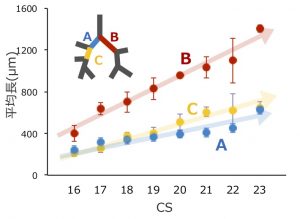
第73回日本人類学会で発表しました(2019.10.12-14、佐賀)。

われわれの研究も人類の研究なので、発表してみました。どの程度受け入れられたでしょうか。

ヒト胚子期における気管支分岐形成の三次元的定量解析
藤井 瀬菜、村中 太河、松林 潤、米山 明男、兵藤 一行、山田 重人、高桑 徹也
ヒト胚子期・胎児期初期における気管・軟骨の3次元的解析
山崎 優、藤井 瀬菜、石川 葵、山田 重人、高桑 徹也
Carnegie Stage (CS) でのヒト後腎における腎小体の発生段階と立体的分布
北沢 遥、石山 華、石川 葵、松林 潤、山田 重人、高桑 徹也

TMIMS symposiumで発表

大脳subplate層の形成

20th TMIMS International Symposium “Principles of Neocortical Development and Evolution” (2019.7.30, Tokyo)で発表しました

Terashima M, Ishikawa A, Yamada S, Takakuwa T, Morphogenesis of the layer structure of cerebral cortex during human late-embryonic period

第59回先天異常学会で発表

第59回先天異常学会で、発表しました(2019.7.26-28, 名古屋)The 13th World Congress of the International Cleft Lip and Palate Foundation -CLEFT 2019-と合同開催でした。

Nohara A, Owaki N, Manesco C, Katsube M, Yamada S, Imai H, Matsuda T, Yoneyama A, Takakuwa T, Relationship between fusion of lateral palatal shelves and growth of Mandible (Meckel’s cartilage)

PUH occurred independently of liver

Takakuwa T. Intestinal loop formation: herniation into the extraembryonic coelom and return to the abdominal coelom (招待講演)

Drastic changes occur during the development of the intestinal loop (IL), including physiological umbilical herniation (PUH) and its return. The present study was designed to analyze such developments three-dimensionally during human embryonic and early fetal period.

Materials and Methods: The software AMIRA was used to analyze the 3D digitalized data (high-resolution MRI, phase-contrast X-ray CT) obtained from the Kyoto Collection.

Results and Discussion: Based on the results of our analysis, the following time line and main features of IL formation were revealed:

Herniate phase (Carnegie stage (CS)14-CS23, Crown-rump length (CRL) < 35 mm): IL rotation was initially observed as a slight deviation of the duodenum and colorectum from the median plane up to CS16. The PUH was noticeable after CS16. The IL displayed a hairpin-like structure, with the superior mesenteric artery (SMA) running parallel to the straight part and the cecum located to the left at CS18. The IL rotated around the SMA only during the early stages (until CS19). The IL gradually moved away, running transversely after CS19. Embryos with liver malformation showed PUH, which indicated that PUH occurred independent of liver volume.

Transition phase (CRL = 37, 41, and 43 mm): Intestinal return began from proximal to distal part in samples with CRL of 37 mm. The cecum returned before the distal end of the small intestine (ileum) in samples with CRLs of 41 and 43 mm.

Return phase: The cecum immediately reached its final position in the right lower quadrant of the abdomen (the adult position). The anti-clockwise “en-bloc rotation” described by descent and fixation of the cecum in the abdominal cavity may not exist. A rapid increase in the space available for the intestine in the abdominal coelom that exceeded the intestinal volume in the extraembryonic coelom was observed. The height of the umbilical ring increased in a stepwise manner between the transition and return phases and its height in the return phase was comparable to or higher than that of the hernia tip during the herniation phase. We speculated that the space is generated to accommodate the herniated portion of the intestine, similar to the intestine wrapping into the abdominal coelom as the height of the umbilical ring increases.

Conclusion: The data obtained in the present study demonstrate the precise timeline of IL formations, which indicate several points of discrepancy in the results of previous studies.