

カテゴリー: 活動記録
第9回京都府細胞診ワークショップ、実技講習会[7月5日]
第9回京都府細胞診ワークショップ、実技講習会 (京都府臨床検査技師会会員、京都臨床細胞学会会員向け) が平成27年7月5日(日)に行われます。 細胞検査士の資格取得をめざす臨床検査技師を対象に細胞診の作成技術、検鏡につい […]
多元計算解剖学symposiumで発表
平成27年度 新学術領域研究「多元計算解剖学」シンポジウム (2015.4.30:東京大学)で発表しました。 ヒト器官形成期において分岐構造を有する器官の3次元分枝パターンを解析する
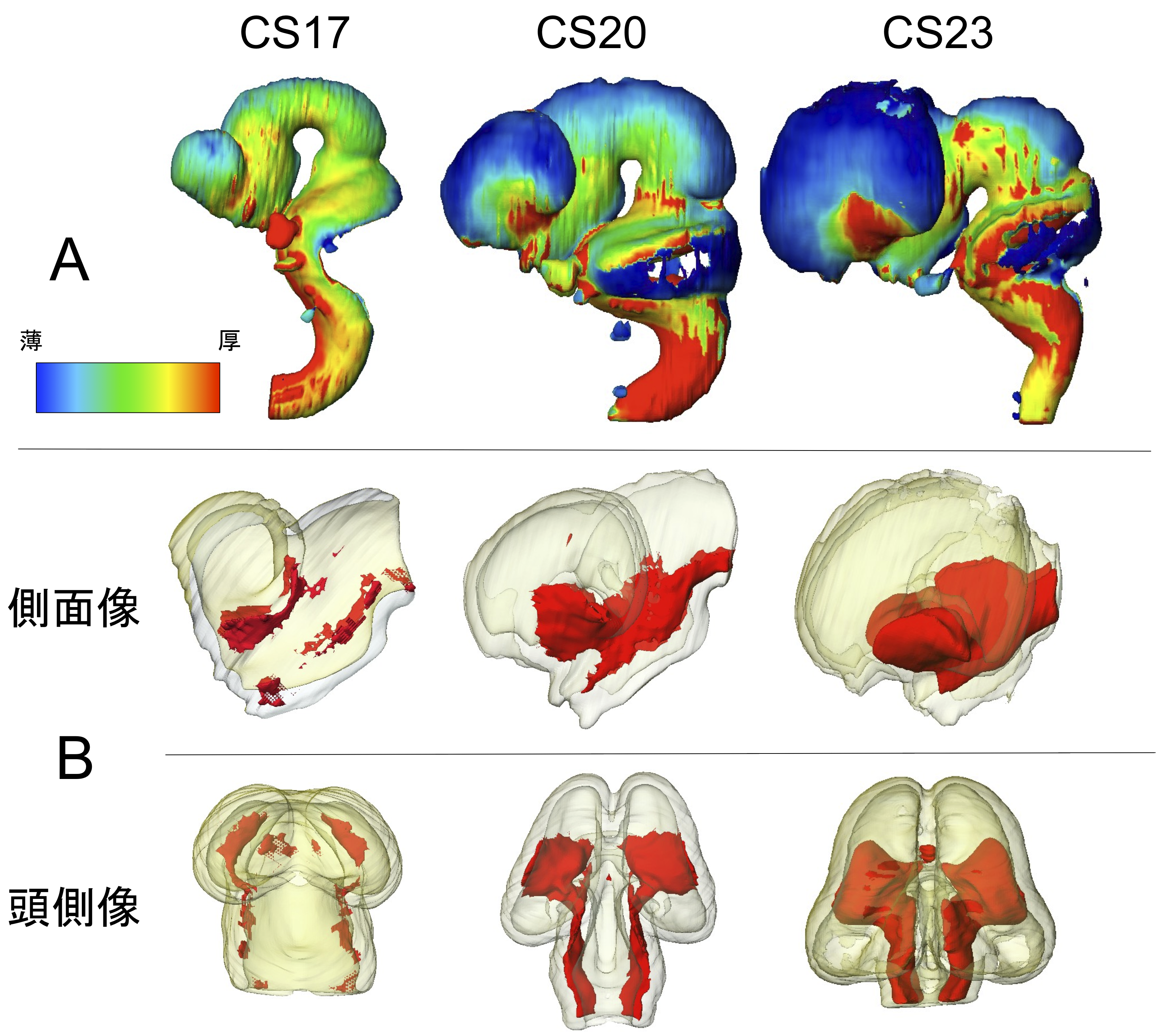
ヒト胚子期における脳形態形成の解析, NeuroImage, Data in Briefに掲載
ヒト胚子期の脳の三次元形成を提示
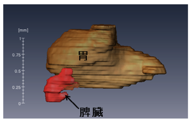
脾臓の形態形成(遠藤卒論)Antat Recに掲載
脾臓の形態形成、内外の血管の形成過程について記載
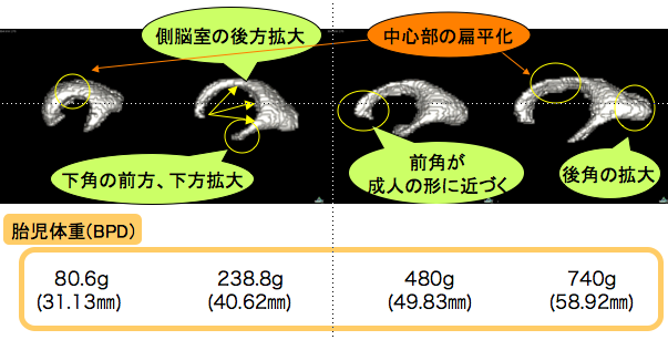
胎児側脳室の形態と長さ計測の有用性(竹谷卒論) Congenit Anomに掲載
妊娠中期ヒト胎児の側脳室の形成を解析
第5回放射光イメージング研究会
第5回放射光イメージング研究会 (3/20, 東京)で金橋くん(研究協力員)が発表しました。 ヒト胚子期における肝臓形態形成異常の解析 位相CT等の放射光を用いて様々なイメージングを行っている方々の集まりです。 &nbs […]

日本科学未来館にヒト胚子立体像が常設展示
3/20から開設された日本科学未来館(東京都江東区)「生命」コーナーにヒト胚子立体像が常設されました(5年間の予定)上記展示に監修者として、協力しました。 新規展示、「細胞たち研究開発中」では、iPS細胞の発見にはじまっ […]

基盤(S)のmeetingに参加
次年度から分担研究者として参加する基盤研究(S)ヒト脳の形態形成から行動生成に至る発達のダイナミクスの研究会議に参加しました。分野の異なる先生の集まりで、なかなか面白い会議でした。
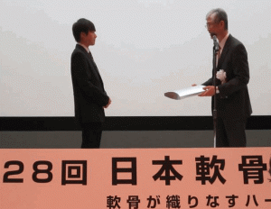
高石くんが優秀演題賞に選出 (28th日本軟骨代謝学会)
「Episcopic fluorescence image captureを用いたラット膝関節腔発生の三次元的解析」高石 亮太、青山 朋樹、張 項凱、樋口 真也、山田 重人、高桑 徹也 (第28回日本軟骨代謝学会 (H2 […]

ヒト胚子透過立体回転画像が知的財産に登録…
ヒト胚子透過立体回転画像が知的財産に登録されました。おもにMRIで撮像されたCS13-23の胚子を、コンピュータを用いて立体化したものです。一部は日本科学未来館で展示される予定です。
修士論文審査が行われました
修士論文審査が行われました。落ち着いてわかりやすく発表でき、良かったと思います。諮問も問題なく終了です。

2014年度;修士論文概要 (尾関)
ヒト聴覚器の各発生段階の形態学的解析 背景:ヒトの聴覚器は、内耳、中耳、外耳に分けられる.その発生過程については、これまで組織切片を用いた観察が主体であり、立体像を用いた解析は膜迷路でしか行われていない.また、現在ヒト発 […]