

カテゴリー: 活動記録
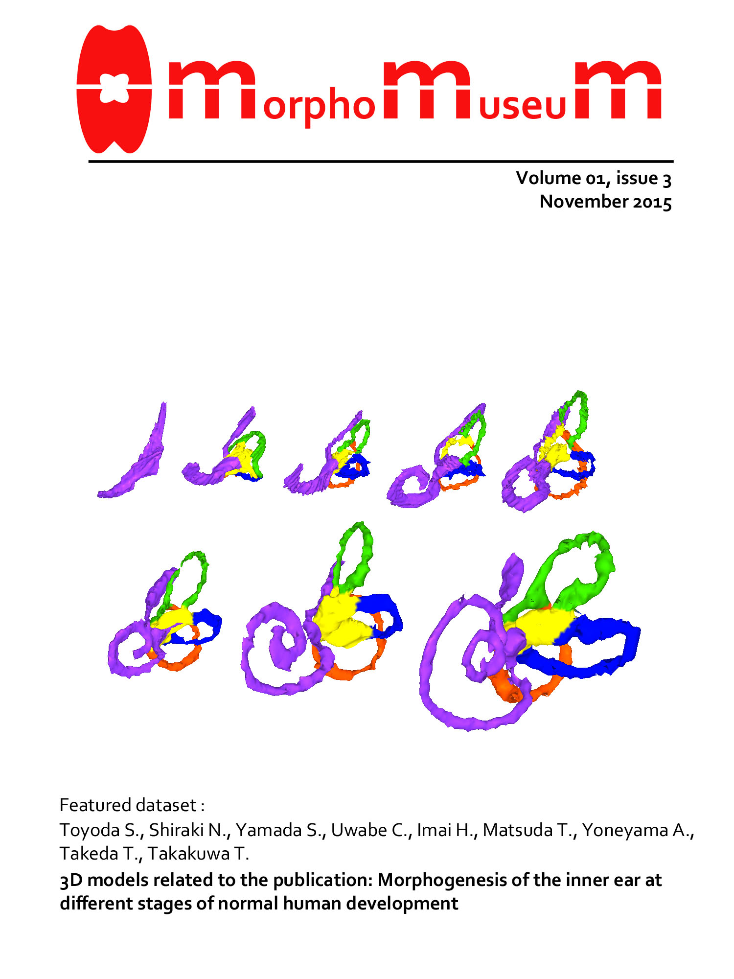
内耳・膜迷路の3D画像が MorphoMuseuMの表紙に採用
ヒト胚子の内耳・膜迷路の3D画像
総説”Human embryology”が発行
ヒトの発生学についての総説が発行されました。New Discoveries in Embryologyという書籍の第5章に収載されています。internetからdownloadできます。 Yamada S, Hill M […]
金橋君の修論がAnat Recに掲載
CS18 -21 の肝形成不全の有病率は、約 1.7%
医用画像研究会(MI)で発表
共同研究者の岸本さん、清水先生ら(東京農工大)が医用画像研究会(MI)で発表されました。 2015.09.08 電気通信大学(調布市) ヒト胚子の眼球を対象とした時空間統計モデルに関する初期検討 岸本 将志、斉藤 篤、大 […]
2015次世代医療を語る -再生医療の実用化に向けて-
今年度の研究科横断型プログラム、次世代医療を語る -再生医療の実用化に向けて-(後期水曜5限)の概要が決定しました。 ※ 京都大学オープンコースウェアに収録済みです。御覧ください。 OCW_2015次世代医療を語る -再 […]
第55回日本先天異常学会で発表
第55回日本先天異常学会学術集会・第38回日本小児遺伝学会学術集会【合同開催】の合同開催で、いつもより盛会でした。 ヒト器官形成期における視覚器の発達について 大坂美穂、山田重人、上部千賀子、米山明男、武田徹、今井宏彦、 […]
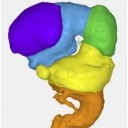
脳の形態形成の3次元データがMorphoMuseuMに掲載
胚子期の脳、脳室の形態像に関する3次元データ

ゲッチンゲン大学に画像取得に行きました
6/10−6/26、ドイツゲッチンゲン大学に共同研究者の山田先生、宮崎さん(山田研)、五十嵐さんの3名が、画像取得に出張いたしました。貴重なBlechshmidt collectionのうちの、連続組織標本をスキャニング […]
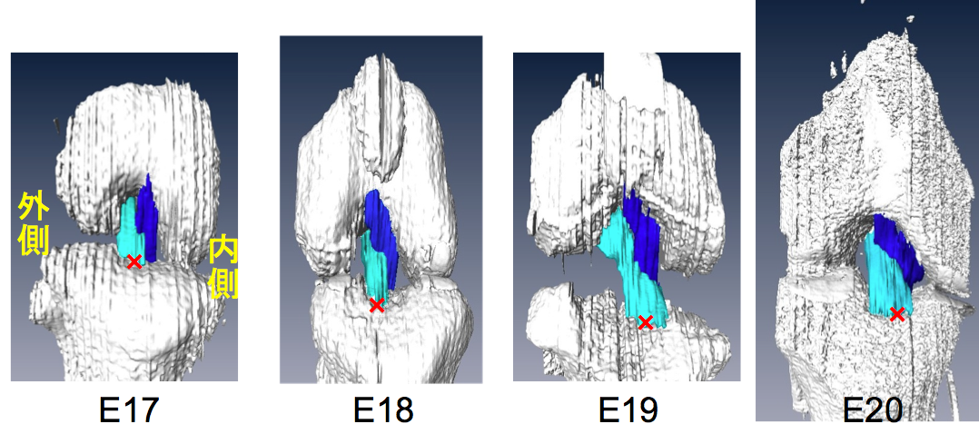
張・高石・樋口君の論文が PlosOneに掲載
ラットの膝関節のACL, PCL靭帯の形成を検討

基盤Sメンバーが来られました。
S基盤研究(S)ヒト脳の形態形成から行動生成に至る発達のダイナミクスのメンバーが東京から来られ、研究についてのDiscussionを行いました。
第104回日本病理学会で発表
第104回日本病理学会総会(4/30-5/2、名古屋)で発表しました。 2年ぶりなので演題をたくさん持って行きました。 「先天異常」というのは病理学の大事な分野の一つなのですが、病理学会内で、私たちと類似のことを行ってい […]
第5回病理学技術者講習会(西日本)[6月27日]
第5回病理学技術者講習会(西日本) が平成27年6月27日(土)に行われます。 臨床検査技師を対象に、病理組織標本作成の技術の向上を目的に開催されるものです。 病理学研究室は、開催に協力することになりました。 >& […]