

カテゴリー: 活動記録
Biological Synchrotron Radiologyで発表
位相CTのヒト発生学への応用について、山田先生、米山先生らが国際学会で発表しました。金橋くんも胚子撮像に参加しており、学会発表の共同演者になりました。 Yamada S, Yoneyama A, Kahanashi T, […]
第53回日本先天異常学会学術集会で発表
2013年7月21日(日)-23日(火)(大阪) ポスター ヒト胚子期における消化管の初期形成と分化 上野沙季 ヒト胚子外表写真を用いたカーネギーステージ(CS)分類判定支援装置の作成 尾関舞美 ヒト胚子期における肝臓の […]
より高解像度の立体情報取得を-PF共同利用実験-
私たちはMR顕微鏡を用いて得られた3次元情報を解析し成果を報告して来ました。そのMR顕微鏡の解像度は最高で35μm/pixel程度で、MRIの技術による撮像の最先端といえます。しかしながら、胚子期の初期や、個々の胚子の器 […]
H25年度”次世代医療を語る”
次世代の医療は、医学研究科だけでなく、さまざまな分野の学生が担って行く可能性があります。今回は、その代表として理工学、細胞生物学、地域・社会学からのアプローチを取り上げ、医療の動向を議論したいと思います。本プログラムを聴 […]
“構成論的発達科学”第2回領域全体会議で発表
構成論的発達科学 第2回領域全体会議̶— 胎児からの発達原理の解明に基づく発達障害のシステム的理解に出席しました (東京大学2013.6.18-19) 発表演題 (京都コレクションと三次元イメージン […]
第102回日本病理学会で発表
第102回日本病理学会(2013.6.6-6.8、札幌)で発表しました。・ 病理学会は2年ぶりの参加でしたので、演題をたくさん持って行きました。 口演 ヒト胚子期における隣接器官による肝臓の形態形成への影響 高桑徹也、山 […]
ヒト胚子模型の作成•体系化
私たちの研究室では、ヒト胚子由来の立体情報を含んだデジタルデータを主に扱っています。それらをもとに形態学的、形態計測学的解析を行い、その成果を学術論文として公表しています。しかしながら、学術論文は、世間一般の方の目に触れ […]
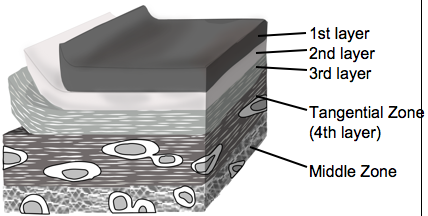
関節の表面は、どのような“層”でできているのか
藤岡さんは修士研究で、関節軟骨の最も表面にある領域が、どのような構造をもっているのかを詳しく解析しました。 関節軟骨は、膝などの関節で骨の表面を覆い、関節をなめらかに動かすために欠かせない組織です。歩く、走る、立つといっ […]
白石君が日本解剖学会symposiumで招待講演
日本解剖学会のシンポジウム「器官形成・発達障害研究におけるMRI定量解析の最先端」で白石くんが「ヒト胚子期の脳形成」の講演(招待)を行いました。 *シンポジストに指名していただきありがとうございました。 ポスター ◆ […]
臨床解剖学実習 (修士)を開講
山田重人教授の全面的協力を得て人体解剖について学ぶ機会を設けました。 よりよい医療の実現のためには解剖学の知識が重要となります。なぜなら、解剖学を基本としてヒトの構造、生理、機能、病理ひいてはヒトそのものの理解へと繋がる […]
新学術領域研究”構成論的発達科学”に参加協力
新学術領域研究(H24-28);構成論的発達科学ー胎児からの発達原理の解明に基づく発達障害のシステム的理解に協力することになりました。研究室の大学院生は臨床検査技師として産科領域のエコーを学びながら研究に参画します。学位 […]

3D解析を応用したヒト発生解剖;年間1500 downloads達成
私たちの研究活動を世界に発信